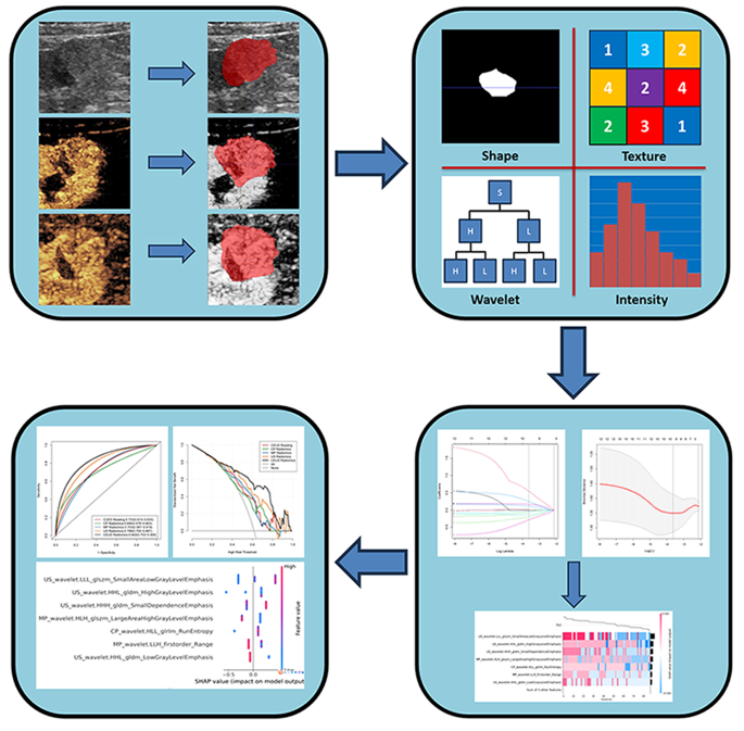
Sufferers and datasets
This analysis was authorized by the Institutional Assessment Committee of the West China Faculty of Public Well being and West China Fourth Hospital, and the requirement for knowledgeable consent was waived. CEUS pictures of fully resected ccRCCs from December 2017 to January 2024 on the West China Faculty of Public Well being, West China Fourth Hospital, and Nanchong Central Hospital have been used on this examine. A complete of 122 ccRCC sufferers have been obtained from the picture databases of the 2 hospitals to type a coaching and testing set in a 7:3 ratio. The inclusion standards have been: (1) ccRCC sufferers who underwent partial or radical nephrectomy. (2) Sufferers who underwent CEUS examination inside two weeks earlier than surgical procedure, (3) sufferers with full scientific knowledge, and (4) no earlier renal surgical procedure or different therapy carried out on suspected ccRCC lesions. The exclusion standards have been as follows: (1) sufferers who underwent anticancer remedy (resembling radiotherapy, chemotherapy, and ablation) earlier than CEUS examination; (2) sufferers with a historical past of each kidney tumors and different sorts of tumors; and (3) sufferers with CEUS picture loss or poor picture high quality.
CEUS picture acquisition
The US devices used to amass the pictures on this examine included IU22 and EPIQ7 (Philips, Amsterdam, the Netherlands) and GE LOGIQ E9 (Common Electrical Co., USA). First, grayscale ultrasound was used to look at the higher stomach, and the scanning sound window, depth, acquire, dynamic vary, mechanical index, output energy, and focal space of the mass have been adjusted to acquire the optimum CEUS picture. Ultrasound distinction brokers (SonoVue; Bracco, Italy) have been used, and 1.5 ml of the distinction agent was injected by way of the elbow vein. Subsequently, the cells have been washed with 5 ml of physiological saline resolution. The timer started counting after the injection of the distinction agent. A low mechanical index (MI < 0.1) was used for the CEUS examination. In line with the rules, CEUS examination is split right into a renal cortical section (CP) 15–30 s after UCA administration with scientific enhancement seen and a renal medullary section(MP), the place each cortical enhancement and medullary enhancement happen 25s–4 min after UCA administration [8]. Grey-scale pictures of the affected person’s most vital space, cortical tumor picture, and medullary tumor picture for evaluation.
Pathological analysis
Two pathologists evaluated all circumstances by observing hematoxylin and eosin (HE)-stained sections below a microscope. All circumstances have been labeled in response to the requirements of the 2016 WHO/ISUP grading system [4]. Divide ccRCC tumors into low-grade (grades I and II) and high-grade (grades III and IV) teams in response to the 2016 WHO/ISUP grading system [4]. If there was a distinction in opinion, one other senior pathologist was invited to take part within the dialogue and attain a consensus.
CEUS evaluation
Ultrasound examination was retrospectively evaluated by two knowledgeable radiologists (engaged in CEUS work for roughly 9 and 5 years) on ultrasound distinction pictures of all sufferers unaware of the pathological and scientific outcomes. Two reviewers independently evaluated the next imaging options of every ccRCC: (a) dimension; (b) echogenicity(Hyper/Iso/Hypo); (c) form(irregular/common); (d) margin (unclear/clear); (e) CEUS improve velocity (when the enhancement depth of cortical tumors is bigger than or equal to the encompassing renal parenchyma, it’s labeled as “quick”; in any other case, it’s thought of as “gradual”); (f) washout (comparability of enhancement depth and renal cortical echo depth in medullary stage tumors); (g) the pseudocapsule(sure/no); (h) necrosis(sure/no). If there was a distinction within the CEUS outcomes of the affected person, the 2 individuals have been evaluated after dialogue.
Picture segmentation and ultrasound radiomics characteristic extraction
We took the next steps within the preprocessing course of. First, we standardized the picture grayscale to make sure consistency within the grayscale distribution within the picture. Subsequent, we adopted image-denoising methods to cut back the attainable interference of noise throughout characteristic extraction. As well as, we resampled the pictures utilizing a linear interpolation algorithm to acquire a standardized voxel spacing of 1 × 1 × 1 mm (x, y, z). We used ITK software program(3.8.0, assist://www.itksnap.org/pmwiki/pmwiki.php? n=Downloads.SNAP3)for handbook segmentation, a broadly used instrument in medical picture processing. The handbook segmentation course of is just not restricted to traditional two-dimensional ultrasound pictures however consists of enhanced ultrasound cortical section pictures and ultrasound medullary section pictures. The world of curiosity refers back to the whole lesion space. In the course of the delineation course of, particular consideration is paid to make sure that the realm of curiosity totally covers the lesion to acquire correct lesion boundaries. As well as, to cut back errors, all handbook segmentations have been independently carried out by two skilled radiologists (with 10 and seven years of expertise in stomach ultrasound analysis), and discussions and negotiations have been performed as wanted to achieve a consensus on the segmentation outcomes. We used intra- and inter-class correlation coefficients (ICC) to judge characteristic stability. Particularly, we randomly chosen renal ultrasound pictures from 50 sufferers and had two radiologists calibrate their respective areas of curiosity (ROIs). Subsequently, Radiologist 1 repeated the identical steps two weeks later and extracted the imaging omics options. We evaluated the consistency and stability of the characteristic extraction by calculating the intra- and inter-group correlation coefficients of those three units of options. To make sure the reliability of the outcomes, we set a threshold for ICC values larger than 0.75, indicating that these options have good consistency and stability and are appropriate for subsequent quantitative evaluation. The remaining pictures have been independently ROI-segmented by radiologist 1, and solely options with good correlation and stability have been retained for subsequent analyses. This technique is adopted to make sure the evaluation’s accuracy and reliability whereas avoiding potential errors brought on by segmentation inconsistency or characteristic instability. Utilizing PyRadiomics (model 3.0.1, https://github.com/AIM-Harvard/pyradiomics.)extracting radiomics options from ultrasound pictures. Applied quite a few engineering-coding characteristic algorithms. The steps for screening ultrasound omics options and setting up ultrasound omics fashions are proven in Fig. 1: Firstly, the options with ICC > 0.75 within the coaching set have been retained. Second, a single-factor rank-sum take a look at was used to display for statistically vital characteristic variations between the coaching set’s high- and low-grade teams. Subsequently, Pearson’s correlation evaluation was carried out on the remaining radiomics options to take away extremely correlated options. Lastly, the LASSO algorithm is used to pick out the optimum options, and the Xgboost algorithm is used to ascertain a radiomics mannequin.
To beat the “black field” nature of ML fashions, the SHAP technique [17] explains every variable’s affect on the optimum efficiency mannequin. The SHAP technique relies on alliance sport idea and calculates the SHAP worth, which evaluates every variable’s marginal contribution to the mannequin’s closing prediction. A number of circumstances have been proposed to disclose how one of the best mannequin generated every prediction. As well as, the SHAP values for every variable in all sufferers have been summarized and averaged to acquire a queue view for international interpretation.
Statistical evaluation
All statistical analyses have been performed utilizing SPSS (model 25.0; IBM Corp., Armonk, NY, USA) and Python 2.7 (Python Software program Basis, Beaverton, OR, USA). Quantitative knowledge with regular distribution is represented as commonplace deviation. Categorical variables are expressed as numbers and percentages. The chi-square take a look at, two unbiased pattern t-tests, and Mann-Whitney U-test have been used for univariate evaluation. Logistic regression evaluation carried out univariate and multivariate analyses of the scientific parameters. Statistical significance was set at p < 0.05. vital.
The receiver working attribute(ROC) curve, space below the curve (AUC), and determination curve evaluation(DCA) have been used to judge the predictive efficiency, calibration capability, and scientific practicality of the fashions. Different distinguishing indicators included accuracy, sensitivity, specificity, constructive predictive worth(PPV), and detrimental predictive worth(NPV).


