A newly developed contrast-enhanced ultrasound (CEUS) Vesical Imaging-Reporting and Knowledge System (VI-RADS) helps each novice and skilled readers to judge muscle-invasive bladder most cancers (MIBC), researchers reported September 10 in Radiology.
“Distinction-enhanced ultrasound (CEUS) with microbubbles makes it potential to obviously determine and assess the three-layer construction of bladder wall by way of real-time perfusion imaging,” famous a crew led by Jing Han, MD, of the Guangdong Provincial Scientific Analysis Heart for Most cancers in Guangzhou, China, which launched the CEUS VI-RADS system.
Whereas CEUS is just not used clinically for this indication as generally as multiparametric MRI (mpMRI), it affords a chance to judge muscle invasion preoperatively. Han and colleagues defined that mpMRI is probably not appropriate for individuals with contraindications, reminiscent of metallic implants and claustrophobia. Importantly, “in settings the place mpMRI is just not available or is contraindicated, CEUS might function a substitute for consider muscle invasion in bladder most cancers,” they wrote, “however mustn’t change mpMRI totally within the analysis of bladder most cancers.”
Utilizing options derived from contrast-enhanced ultrasound pictures acquired by way of transabdominal and intracavity approaches, the crew developed and validated the CEUS VI-RADS metric. The research enrolled 193 sufferers with suspected bladder most cancers who underwent transabdominal or intracavity CEUS between July 2021 and Might 2023; contributors have been divided into a coaching set of 126 and a validation set of 67.
Within the coaching set, 9 options related to MIBC at ultrasound and CEUS have been recognized. Amongst them, continuity of hyperechoic interior layer, interruption of hypoechoic muscularis propria, and extension of suspicious tumor echo to extravesical fats have been unable to be evaluated on 15% (19 of 126), 28% (35 of 126), and 15% (19 of 126) of ultrasound examinations, respectively, and have been thus excluded from the CEUS VI-RADS.
Imaging options deemed main included lesion measurement, vascular stalk at CEUS, early hyper-enhanced steady interior layer, well-defined steady layer of hypo-enhanced muscularis propria, tumor early enhancement of muscularis propria, and extension of tumor early enhancement to serosa and extravesical fats.
The researchers reported the likelihood of muscle invasion utilizing CEUS VI-RADS as the next:
- 0% CEUS VI-RADS 1
- 2% CEUS VI-RADS 2
- 11% CEUS VI-RADS 3
- 53% CEUS VI-RADS 4
- 100% CEUS VI-RADS 5
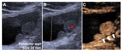
 Distinction-enhanced ultrasound (CEUS) Vesical Imaging-Reporting and Knowledge System (VI-RADS) class 3 findings in a 40-year-old male with non-muscle-invasive high-grade urothelial carcinoma. (A) Grayscale US picture obtained utilizing a convex array probe (5C1) by a transabdominal strategy confirmed a 35-mm lesion on the bladder posterior wall. (B) Colour Doppler US picture confirmed interior vascularity throughout the lesion. (C) Distinction-enhanced US picture confirmed well-defined steady layer of hypo-enhanced muscularis propria (white arrows) with out vascular stalk on contrast-enhanced US or early hyper-enhanced steady interior layer. The remark was assigned to VI-RADS 3 based mostly on contrast-enhanced US by the readers devising the CEUS VI-RADS. Picture and caption courtesy of the RSNA.
The analysis is essential as CEUS has the potential to depict MIBC sooner and at a decrease price, based on Glen Morrell, MD, PhD, of the College of Utah in Salt Lake Metropolis, who wrote a commentary that accompanied the research.
“In settings the place MRI is just not out there, ultrasound could be a invaluable various,” Morrell wrote. “Nevertheless, CEUS does have some relative disadvantages, which might restrict its use … CEUS requires the number of a single picture aircraft earlier than injection of distinction materials. This introduces the danger of lacking muscle-invasive tumor at CEUS by undersampling. Moreover, CEUS is extra invasive than MRI, requiring using intracavitary transrectal or transvaginal US probes for optimum depiction of bladder tumors.”
The whole research might be discovered right here.

