Sufferers
The Xiangyang Metropolis Centre Hospital’s institutional evaluation board authorized this retrospective investigation and eliminated the demand for knowledgeable consent.
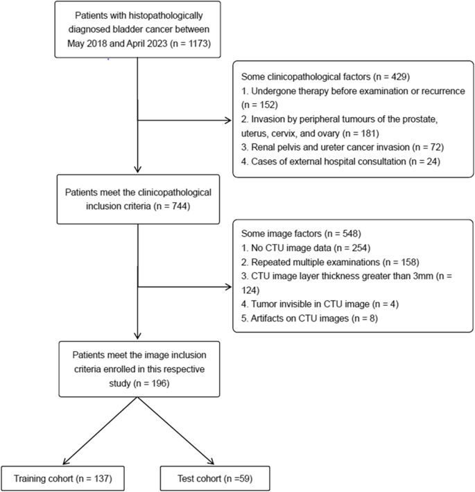
We searched our hospital database for sufferers with BCa confirmed by postoperative pathology from Could 2018 to April 2023. These standards had been used to find out inclusion: [1] sufferers who underwent TURBT or radical cystectomy with pathologically confirmed urothelial carcinoma, and [2] sufferers who underwent CT urography (CTU) inside the preoperative interval. The next standards had been used to exclude sufferers: [1] artifacts on CTU photos, [2] incomplete picture sequence and/or layer thickness better than 3 mm, [3] lesion width decrease than 5 mm, [4] underdistended bladder, [5] biopsy or therapy comparable to chemotherapy or radiotherapy previous to the CT examination, and [6] muscle layer undistinguishable in specimens resected by TURBT. A complete of 196 sufferers had been included, together with 97 NMIBC and 89 MIBC instances, as proven in Fig. 1. Instances had been divided into coaching and take a look at cohorts (n = 137, n = 59) by 7:3 stratified random sampling.
The affected person’s demographic and clinicopathological info was retrieved from their medical document, together with age, intercourse, historical past of smoking, medical grievance (e.g., hematuria, hydronephrosis, or incidental findings), infiltrative standing of the bladder wall’s muscular layer, and histopathological grading (excessive and low grades as decided by utilizing biopsy previous to surgical procedure). Furthermore, imaging info was retrieved from the affected person’s CT photos, and the variety of lesions, calcifications, lesion size and tortuous blood vessels seen to the bare eye round or inside the lesions had been documented (Supplementary Materials S1).
CT information and picture acquisition
All enrolled sufferers underwent CTU inside 1–2 weeks prior surgical procedure. Earlier than the scan, sufferers had been instructed to drink between 800 and 1000 ml of water, however to not urinate till the scan was over. After scanning, 50 mL of ioversol or 100 mL of iopamidol had been intravenously administered, adopted by 50 mL of saline at a price of three mL/s. Photographs of the renal corticomedullary, nephrographic, and excretory part had been obtained at 25 s, 75 s, and 300 s after the thresholding of the thoracoabdominal aortic junction was reached. Subsequent analyses used solely axial nephrographic part photos. Multidetector CT scanners with 64 to 128 detector rows (Siemens Healthineers, Philips Brilliance, and Philips Brilliance iCT) had been used to acquire CT photos. The scanning parameters had been 120 kV, computerized mA settings (vary 80–320 mA), layer spacing of 1 mm, and layer thickness of 1.5–3 mm. Smooth tissue algorithm (window width (WW): 300–500 HU, window stage (WL): 45–60 HU) had been used after imaging.
CT picture segmentation and have extraction
Three-dimensional areas of curiosity (3D-ROIs) had been manually outlined on thin-layer CT photos in the course of the nephrography part utilizing the ITK-SNAP program (model 4.0.1; http://itk-snap.org). Solely the biggest lesion in sufferers who had a number of lesions was chosen for segmentation on this research. 3D-ROIs alongside the tumor margins had been manually drawn by radiologist 1 (ZR, with 6 years of genitourinary imaging expertise and 5 years of tumor segmentation expertise). Radiologist 1 was uninformed of the standing of muscle invasion in its postoperative pathology. To make sure the reproducibility of the areas of curiosity (ROI), intragroup correlation (ICC) was used to evaluate intra-observer settlement. We randomly chosen 30 sufferers, and the ROI was manually outlined once more after 4 weeks by the identical radiologist and one other one (ZLH, with 3 years of expertise in genitourinary imaging and tumor segmentation). A superb settlement was outlined as an ICC of 0.75 or larger.
The PyRadiomics bundle (model 3.0.1, obtainable at https://github.com/AIM-Harvard/pyradiomics.git) and Python (model 3.7.5) had been used to acquire the radiomics options from the CT photos. The unique and wavelet-filtered picture allowed for the retrieval of all radiomics options, which may very well be divided into seven teams, particularly first-order statistics, form options, glcm options, gldm options, glrlm options, glszm options and ngtdm options. The options extraction methodology is out there from https://pyradiomics.readthedocs.io/en/newest. Lastly, 851 options had been extracted in every quantity of curiosity of CT photos, respectively.
Function choice and Radiomics mannequin constructing
All options had been normalized utilizing z-score normalization previous to characteristic choice and mannequin improvement. The ICC was used to evaluate the repeatability of every radiomics characteristic each intra- and inter-observer. In our research, solely the options with ICC values better than 0.75 had been included. Options filtering was carried out by utilizing a significance take a look at (Scholar’s t-test if the info adhered to a standard distribution, in any other case the Mann-Whitney U take a look at) to pick the options with excessive predictive energy (p < 0.05). Probably the most helpful predictive muscle invasion status-related options had been then chosen from the coaching cohort utilizing the least absolute shrinkage and choice operator (LASSO) and 10-fold cross-validations. The radiomics rating (Radscore) was obtained by making use of linear weighting primarily based on the chosen options by the LASSO algorithm.
Growth and efficiency of a Radiomics-clinical nomogram
The included medical indicators had been subjected to unbiased univariate and multivariate analyses to determine the clinicopathological predictors for muscle invasion and create a medical mannequin. The ultimate predictors for muscle invasion had been chosen by utilizing a multivariate logistic regression evaluation that included the Radscores and the unbiased medical indicators. The method used ahead stepdown choice with a liberal p < 0.05 because the retention standards. Based mostly on the outcomes of the multivariate logistic regression evaluation, a radiomics-clinical nomogram was created. Using the Hosmer-Lemeshow take a look at and the calibration curve, the radiomics-clinical nomogram’s calibration was evaluated.
Fashions comparability and medical usefulness analysis
To additional assess the applicability of the radiomics-clinical nomogram, it was in contrast with the medical and the radiomics mannequin. The diagnostic efficacy of the totally different fashions for BCa prediction was assessed by utilizing receiver working attribute (ROC) curve to quantify the evaluation energy of every mannequin. The world beneath the ROC curve (AUC) and 95% confidence intervals (CI) together with specificity, sensitivity, accuracy, damaging predictive worth (NPV), constructive predictive worth (PPV) had been used to investigate the diagnostic efficacy of the radiomics fashions. Choice curve evaluation (DCA), which calculated the online profit at totally different threshold possibilities and in contrast the radiomics-clinical nomogram with the medical mannequin, was used to judge the medical worth of the radiomics-clinical nomogram.
Statistics
The statistical evaluation was accomplished utilizing R (model 3.6.1, accessible at https://www.r-project.org). Picture manufacturing was carried out utilizing Microsoft Visio for Home windows (model 2021). Information that failed to adapt to the traditional distribution standards had been in contrast between the 2 teams utilizing the Mann-Whitney U take a look at. Following a normality examine, the continual information had been used the Scholar’s t-test. Regular information had been represented as imply ± commonplace deviation. Counting information had been reported because the variety of instances (price), and the chi-square take a look at was utilized to check the 2 teams. Inter-observer reproducibility of radiomics traits was assessed utilizing the ICC to judge inter-observer settlement between radiologists, with coefficients better than 0.75 indicating good reproducibility. The diagnostic efficacy of the medical and radiomics mannequin, in addition to radiomics-clinical nomogram for BCa prediction had been assessed utilizing ROC curve. To find out the p worth for the AUC, DeLong’s take a look at was utilized. The 2-sided p worth threshold for statistical significance was 0.05.


