Screening mammography exams organized from low to excessive breast density can increase radiologists’ interpretation efficiency, counsel findings printed on October 8 in Radiology.
Researchers led by doctoral candidates Jessie Gommers and Sarah Verboom from Radboud College Medical Middle in Nijmegen, the Netherlands, discovered slight enhancements within the batch studying efficiency of radiologists when exams had been ordered this manner, in addition to decreased studying occasions.
“Though bigger research are wanted to validate these findings, our outcomes counsel that ordering screening mammograms for studying by growing volumetric breast density could also be possible,” the research authors wrote.
There’s a have to strategize round figuring out most cancers in complicated mammographic backgrounds, excessive volumes of screening exams, and the low prevalence of breast cancers at screening, the researchers recommended. This concept stems from earlier research’ findings that between 20% and 50% of interval and screen-detected breast cancers had been seen on prior screening mammograms.
Visible adaptation can impression screening efficiency. For batch studying, this requires decoding radiologists to adapt to the traits of the mammogram they’re studying. Which means that mammograms for interpretation must be ordered in order that photos with traits selling comparable visible adaptation are grouped collectively.
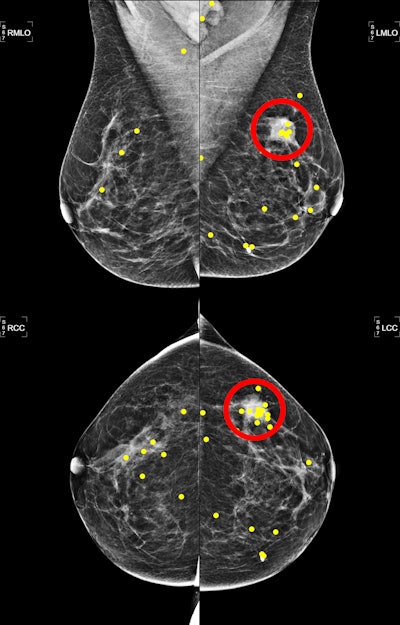
Gommers, Verboom, and colleagues studied how the screening efficiency of radiologists throughout batch studying of screening mammograms could possibly be improved when the exams are ordered in accordance with traits that will promote visible adaptation and fixation occasions (that’s, the time the reader spends on the picture).
 Mammograms depict a 54-year-old girl with an invasive ductal carcinoma within the lateral higher quadrant of the left breast. The yellow dots signify eye fixations, and the pink circle represents the lesion area. Picture courtesy of the RSNA.
The evaluation included screening mammograms carried out between 2016 and 2019 for 150 ladies with a median age of 55 years. Of the mammograms, 75 had breast most cancers whereas the remaining 75 didn’t. The screening exams, every consisting of 4 mammograms, had been interpreted by 13 radiologists in three distinct orders: randomly, by growing volumetric breast density, and primarily based on a self-supervised studying encoding. The researchers additionally employed an eye fixed tracker to report eye actions by the radiologists.
Exams ordered by growing breast density achieved the very best marks of the three methods, together with having increased space underneath the curve (AUC) values and discount in studying and fixation occasions.
| Outcomes of batch studying ordering methods | |||
|---|---|---|---|
| Measure | Random | Self-supervised studying | Volumetric breast density |
| AUC | 0.92 | 0.92 | 0.93 |
| Sensitivity | 81% | 80% | 81% |
| Specificity | 86% | 84% | 89% |
| Studying time | 27.9 seconds | 28.4 seconds | 24.3 seconds |
| Fixation time | 4.6 seconds | 4.6 seconds | 3.7 seconds |
| Fixation rely | 52 | 54 | 47 |
| *Volumetric breast density technique achieved statistical significance in comparison with random ordering for all measures apart from sensitivity. Random ordering achieved statistical significance in comparison with self-supervised studying for fixation rely and studying time. | |||
The research authors recommended that automated density algorithms may assist arrange screening program worklists and additional enhance the flexibility of radiologists to detect and characterize suspicious mammographic findings.
“Nonetheless, future analysis ought to assess what different order standards could additional optimize radiologists’ screening efficiency,” they added. “If confirmed profitable, future research also needs to assess the effectiveness of those ordering methods prospectively in precise screening settings.”
In an accompanying editorial, Lars Grimm, MD, from Duke College in Durham, NC, wrote that the research’s outcomes are promising.
“Though vital extra testing and refinement is required, the findings signify a uncommon alternative to enhance breast radiologists’ interpretation efficiency and effectivity and are complementary to current efficiency enchancment measures, together with batch studying and newer AI instruments,” Grimm wrote.
The total research might be discovered right here.

