Dataset
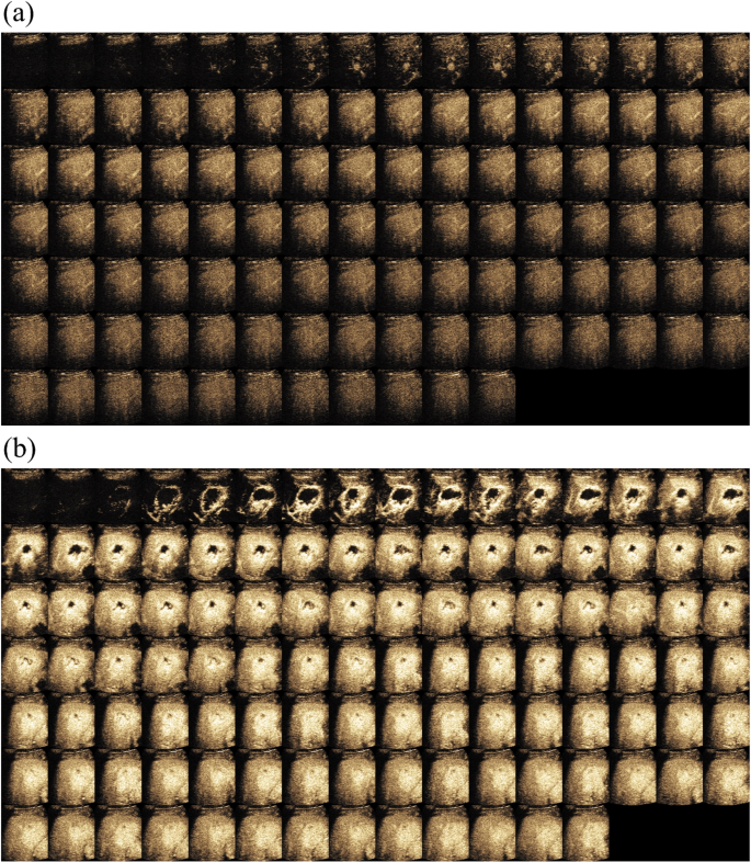
It is a retrospective research and was authorized by the Institutional Evaluation Board and Ethics Committee. The requirement for knowledgeable consent was waived off. From Might 2018 to Might 2019, 440 sufferers with focal liver lesion who underwent CEUS examination have been recruited. The CEUS examinations have been taken utilizing completely different ultrasound imaging programs (Philips, Siemens, Mindray and Canon medical programs). In the course of the examination, a bolus of 1.5-2.4 ml of Sonovue (Bracco, Milan, Italy) was injected intravenously by way of a cubital vein, adopted by a flush of NaCl 0.9% 5 ml in bolus. Actual-time aspect by aspect contrast-enhanced mode was turned on with the mechanical index (MI) was set as little as attainable at vary of 0.05 to 0.1. Normally, clinicians file a number of loops from the top of distinction agent injection to about 6 minutes. All CEUS loops have been saved in DICOM format. Every body in loop had each CEUS information and B-mode information in twin show with decision in 960 (Peak) x 1280 (Width) x 3 (RGB). Given this was an retrospective research, the imaging parameter settings on completely different ultrasound programs weren’t essentially the identical. Ultrasound docs adjusted the parameters whereas scanning to acquire the optimum picture high quality for them to make the prognosis. That is extra near frequent state of affairs throughout typical scientific apply. In Fig. 1, we confirmed two typical circumstances in CEUS.
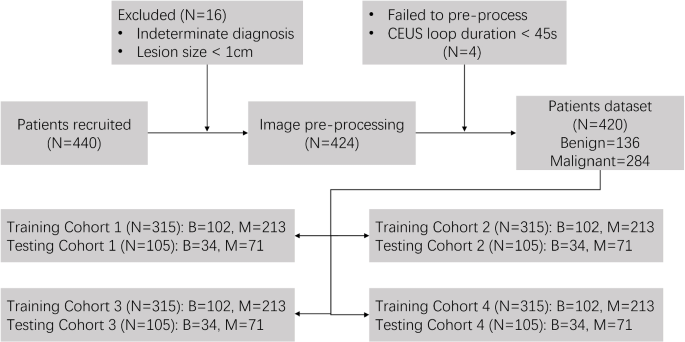
From the 440 sufferers above, we additional chosen with inclusion and exclusion standards as beneath. Inclusion standards have been as follows: 1) aged 18-80 years previous; 2) prognosis confirmed by histologic examination, EASL tips, distinction MRI scanning, or follow-up past 6 months; 3) CEUS loops have been accomplished within the sense that every one three phases together with AP, PVP and LP have been recorded. Exclusion standards have been as follows: 1) lesion dimension (le) 1cm; 2) lack of dicom header or fail to load and preprocess; 3) Length of the primary CEUS loop is shorter than 45s. Lastly, 420 sufferers together with 136 benign and 284 malignant circumstances have been enrolled in our research, proven as Fig. 2. Desk 1 signifies the quantities of benign and malignant circumstances with their corresponding subtypes. Sufferers have been randomly divided into coaching and testing cohorts with 3:1 ratio (4-fold cross-validation) for algorithm growth and analysis.
Methodology
On this part, we’ll introduce the proposed methodology and two strategies for comparability.
Proposed methodology
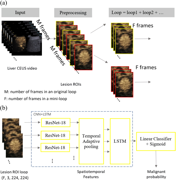
Probably the most intuitive method to make use of deep studying approach within the prognosis of liver lesion with CEUS is to feed the CEUS loops right into a 3D CNN mannequin as a result of 3D CNN preserves each spatial and temporal options. Nevertheless, the 3D CNN has excessive computational complexity. With restricted variety of datasets, the mannequin could be simply overfitted. To beat this challenge, we proposed to make use of the CNN-LSTM mannequin to robotically make the differential prognosis of liver lesion primarily based on CEUS loops. On this methodology, CNN extracts spatial options of every body, adopted by the LSTM to study the temporal options from the sequences of those spatial options. The flowchart of the tactic is proven in Fig. 3 and the detailed description are as follows.
Crop Area of Curiosity Sequence (ROIs) of liver lesion.
Usually in scientific apply, the clinician would label 4 markers to indicate lengthy and brief axis of lesion in a specific body, which could be utilized because the bounding field of lesion. To cut back the impact of respiratory movement, the dimensions of this bounding field is expanded by 50%.
Break up complete CEUS cine-loops into a number of mini-loops.
As talked about, an professional clinician may give dependable and correct prognosis outcomes for liver lesions primarily primarily based on typical CEUS frames from AP, PVP and LP. Leveraging this area information, we chosen F (e.g. F=4) frames at equal time intervals from every part (AP, PVP, LP) to cut back the influence from out of airplane frames which have been brought on by respiratory movement. The removing work was automated with no visible inspection or guide interplay required from a radiologist. It was finished by way of a TIC curve becoming primarily based methodology. For a given body, if absolutely the distinction between its common depth and the TIC fitted depth was bigger than 2 instances of the usual deviation of the typical depth of all frames, it was eliminated. For the reason that period of three phases aren’t the identical, completely different variety of F-frame could also be generated. As urged by tips akin to CEUS LI-RADS [23], the previous couple of seconds of PVP and LP are crucial frames. With this prior data, we choose F frames from again to entrance on the timeline of PVP and LP. Lastly, your entire CEUS loop from every affected person was cut up into a number of mini-loops and every of which has a malignancy chance because the output of the proposed community. The ultimate prediction result’s generated by the bulk vote of mini-loops. In contrast with utilizing the entire loop straight, splitting every CEUS loop into a number of mini-loops can enhance pattern dimension and mitigate the chance of overfitting. This may enhance the full variety of CEUS loops inputting into the community, which in precept equal to augument the info temporally.
Map every mini-loop to malignant chance with CNN-LSTM mannequin.
We employed residual networks due to its good efficiency and ease. Every ROI picture feeds to the ResNet-18 [24] community (ResNet) to extract spatial options. The ResNet18 was pretrained primarily based on ImageNet by freezing the primary 5 layers. Then the F*3 dimensional spatial options of mini-loop are smoothed to be 3 dimensional by way of temporal adaptive pooling with the intention to mimic the options from AP, PVP and LP and to cut back the respiratory movement on the identical time. Afterwards, 3 dimensional spatial options are fed to the LSTM [25] to extract spatio-temporal options by way of adaptive common pooling layer. The LSTM comprises two layers. The primary layer is bidirectional LTSM to have sequence data in each instructions backwards and forwards. The variety of options within the hidden state is 128. Lastly spatio-temporal options from each mini-loop are used to foretell malignancy chance.
Strategies for comparability
On this paper, we in contrast the proposed methodology with a 3D-CNN primarily based methodology and a classical ML-based TIC methodology.
3D-CNN primarily based methodology.
As talked about, probably the most intuitive method to course of video primarily based information is to make use of a 3D-CNN. With restricted variety of datasets in addition to reminiscence, one cannot straight enter the CEUS loop right into a 3D-CNN. Thus, the identical pre-processing step as Fig. 3a was used. As an alternative of utilizing CNN+LSTM as Fig. 3b, 3D-CNN was employed on this methodology.
ML-based TIC methodology.
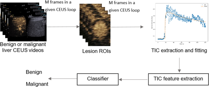
The block diagram of this methodology is proven in Fig. 4. TIC was firstly generated utilizing the sign depth imply worth of a manually drawn ROI inside the lesion. As a consequence of respiratory movement, the TIC is normally very noisy and curve becoming is required. There are 5 completely different perfusion fashions generally used: the lognormal distribution, the gamma variate operate, native density random stroll (LDRW), first passage time (FPT), and the lagged regular operate [26]. In apply, there isn’t a requirements or guideline for selecting one of the best perfusion mannequin. Furthermore, utilizing single perfusion mannequin to estimate perfusion parameters may be very delicate to both the preliminary values for perfusion parameters or the number of applicable boundary circumstances. Due to this fact, we’ve got applied a Multi-Mannequin Framework, which applies nonlinear regression optimizing methodology to all 5 perfusion fashions. Afterwards, the perfusion parameters have been chosen from the mannequin with the bottom estimation error by evaluating varied curve-fitting fashions and deciding on the optimum answer utilizing an exhaustive search approach. This framework was applied in-house in Python. The perfusion parameters embody AP onset time, peak time, peak depth, space beneath the curve, imply transit time, gradient of wash in, gradient of wash out. Lastly, all of those parameters plus the lesion dimension have been enter right into a classifier. XGBoost was used because the classifier and it was chosen primarily based on experiments with greatest outcomes.
On this work, a NVIDA GPU GeForce GTX 1080 Ti 11 GB was adopted to coach completely different fashions. Okay-fold cross-validation (for instance: ok=4 right here) methodology was carried out to keep away from the sampling bias by randomly partitioning dataset stratified by benign and malignant. For the proposed and 3D-CNN primarily based methodology, fashions have been educated by a stochastic gradient descent solver with a cross-entropy loss operate for six epochs in whole, with studying price 0.01 and 0.001 within the first 4 epochs and within the final 2 epochs.
Analysis Metrics
On this research, 4 analysis metrics have been used to judge the mannequin efficiency, specifically: receiver working attribute (ROC) curve, the realm beneath the curve (AUC), sensitivity and specificity. Sensitivity and specificity could be calculated as follows:
$$start{aligned} sensitivity = frac{TP}{TP+FN} finish{aligned}$$
(1)
$$start{aligned} specificity = frac{TN}{TN+FP} finish{aligned}$$
(2)
TP, TN, FP and FN symbolize the variety of true positives, true negatives, false positives and false negatives.


