

Sufferers
This retrospective research was accepted by the institutional assessment board, and the requirement for knowledgeable consent was waived. All sufferers with HCC present process three-phase preoperative DCE-MRI between September 2019 and August 2022 had been enrolled. The inclusion standards had been as follows: (1) pathologically confirmed HCC; (2) singular tumors, with or with out satellite tv for pc nodules—outlined as lesions with a diameter ≤ 2 cm and a distance ≤ 2 cm from the primary tumor; (3) DCE-MRI was carried out inside 1 month earlier than surgical procedure; (4) no prior most cancers remedy, together with transarterial chemoembolization and radiofrequency ablation; and (5) no macrovascular invasion proven on MRI. The exclusion standards had been as follows: (1) particular kinds of liver most cancers reminiscent of double phenotype liver most cancers; (2) poor MRI picture high quality; (3) recurrent HCC; and (4) concurrence of different malignancies. Determine 1 reveals the affected person recruitment course of.
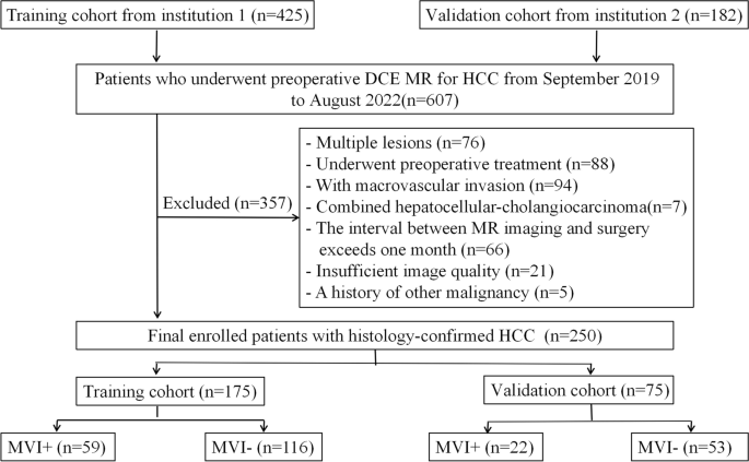
Flowchart of the research sufferers. DCE MRI dynamic contrast-enhanced MRI, HCC hepatocellular carcinoma, MVI microvascular invasion
Laboratory examination and histopathology
Preoperative laboratory indexes (Desk 1) included serum α-fetoprotein (AFP), carcinoembryonic antigen, carbohydrate antigen 199, carbohydrate antigen 125, ferritin, hepatitis B virus, complete bilirubin, direct bilirubin, albumin, and gamma-glutamyl transferase (GGT). The Barcelona Clinic Liver Most cancers (BCLC) staging system was additionally included.
HCC pathological specimens had been collected following the 7-point baseline sampling protocol. Histopathological traits, together with MVI standing and liver fibrosis grade primarily based on the Scheuer scoring system, had been double-blindly decided by two pathologists with greater than 10 years of expertise. MVI means the detection of most cancers cell nests within the vascular lumen lined with endothelial cells underneath a microscope, it’s primarily noticed within the branches of the portal vein.
DCE-MRI protocol
All MR examinations had been carried out with a 3.0 T MRI system (Signa HDXT, GE Medical Techniques, Milwaukee, WI, USA) with intravenous bolus injection of 0.1 mmol/kg gadopentetate dimeglumine (Magnevist®, Bayer Schering Pharma, Berlin, Germany) in each establishments. Scans in a three-dimensional fast-spoiled gradient-recalled echo sequence (liver acceleration quantity acquisition, LAVA) on the arterial, portal vein, and equilibrium phases had been obtained with 20—35 s, 60—90 s, and 160—180 s delays, respectively. The scanning parameters for establishment 1 had been as follows: repetition time, 3.2 ms; echo time, 1.5 ms; reversal angle, 10°; subject of view, 380 × 304 mm; thickness, 2 mm. Whereas the scanning parameters for establishment 2 had been as follows: repetition time, 3.5 ms; echo time, 1.6 ms; reversal angle, 13°; subject of view, 380 × 380 mm; thickness, 2 mm.
Qualitative radiographic descriptors
Picture evaluation was carried out double-blindly by two radiologists with 10 and 15 years of expertise in liver MRI analysis. The next eight imaging traits had been assessed: (a) tumor measurement; (b) tumor gross kind—nodular or non-nodular [14]; (c) rim enhancement within the arterial section [15]; (d) arterial peritumoral parenchymal enhancement [16]; (e) washout [17]; (f) peritumoral hypointensity within the later section [18]; (g) radiological capsule [19]; and (h) intratumoral artery [5].
Tumor segmentation and static radiomics characteristic extraction
The entire tumor was manually depicted together with the lesion define on every axial slice of every MRI section by a radiologist with 10 years of expertise ( reader 1) utilizing ITK-SNAP (http://www.nitrc.org/initiatives/itk-snap/). Moreover, to judge the intra-observer reproducibility and inter-observer reliability of characteristic extraction, photos of 40 sufferers randomly chosen from the coaching cohort after 3 months had been resegmented independently by two radiologist with 10 (reader 1) and seven (reader 2) years of expertise, respectively. The intra-/inter-class correlation coefficients (ICCs) had been used to check the consistency between intra-observer and inter-observer ROIs. For photos with completely different resolutions, all voxel sizes of all photos had been resampled with the identical measurement of 1 × 1 × 1 mm3. The picture gray-scale values had been normalized. The normalization process was primarily based on the next mathematical method:
$$x^{‘}= (x – mu ) /sigma$$
(1)
the place μ is the imply picture density worth, σ is the usual deviation of picture density. The grey values of the photographs had been normalized to 1–64, as really helpful by Orlhac F et al. [20]. Quantitative radiomics parameters had been calculated utilizing MATLAB software program. A complete of 484 radiomics options had been obtained and categorized into three classes, together with 7 depth options, 53 texture options, and 424 wavelet options.
Dynamic radiomics characteristic building
Dynamic radiomics options had been constructed primarily based on the static options change of the identical imaging examination at completely different phases or completely different imaging examinations, which could be expressed as Eq. 2:
$$phi(Psi(x(t1)),Psi(x(t2)),dots,Psi(x(tk)))$$
(2)
the place Ф(.) transforms Rokay to Rd. okay is the section variety of the picture, and d is the variety of extractable dynamic options. The next three kinds of dynamic options had been constructed to replicate the modifications in static options in numerous phases:
-
1.
Built-in options.
The built-in options primarily describe the sample of characteristic modifications with respect to time. Three kinds of built-in options had been studied, together with the imply, variance, and coefficient of variation.
-
2.
Discrete options.
The discrete options primarily describe the sample of characteristic modifications between two consecutive time factors (outlined as a section) and contain two calculation strategies: relative change charge (RCR) and relative common change charge (RACR), that are calculated as in Eqs. 3 and 4
$$RCR(Psi (x(t))) = |Psi (x(tj)) – Psi (x(ti))| / Psi (x(ti)), 1 le j le i le okay$$
(3)
$$RACR(Psi (x(t))) = |Psi (x(tj)) – Psi (x(ti))| / Psi (x(dot{t})), 1 le j le i le okay$$
(4)
On this research, there have been 4 scanning time factors, corresponding to a few segments: plain–arterial section, arterial–portal vein section, and portal vein–equilibrium section. Six kinds of discrete options had been obtained.
-
3.
Parameter becoming options.
Linear, quadratic, and exponential traces had been fitted to the characteristic–time relationships. Parameters of the three becoming strategies, with the utmost curvatures of the quadratic and exponential fittings, had been recorded as dynamic options for the corresponding static characteristic.
The linear becoming equation is expressed as Eq. 5, and okay and d had been extracted as dynamic options.
$$characteristic = okay instances t + d$$
(5)
The quadratic becoming equation is expressed as Eq. 6, and a, b, and c had been extracted as dynamic options.
$$characteristic=atimes t^2+btimes t+c$$
(6)
The curvature of the quadratic operate is expressed as Eq. 7. The time (TmaxQK) comparable to the utmost curvature (maxQOkay) and the curvature (QKmax_feature) comparable to the utmost characteristic worth had been solved for and recorded as two dynamic options.
$$QK=left|2aright|/{(1+left(2atimes t+vibrant)^2)}^{3/2}$$
(7)
The exponential becoming equation is expressed as Eq. 8, and α and β had been obtained as dynamic options.
$$characteristic=alphatimes e^t+beta$$
(8)
The curvature of the exponential operate is expressed as Eq. 9. The time (TmaxEK) comparable to the utmost curvature (maxEK) and the curvature (EKmax_feature) comparable to the utmost characteristic worth had been solved for and recorded as two dynamic options.
$$EK=left|atimes {e}^{t}proper|/{left(1+atimes {e}^{2t}proper)}^{3/2}$$
(9)
A complete of 20 kinds of dynamic radiomics options had been obtained. For every affected person, the variety of static radiomics options was 484, and the variety of dynamic radiomics options was 20 × 484.
Radiomics signature building
The variability between the 2 radiologists’ tumor contours was estimated utilizing ICCs. Steady options with ICCs > 0.8 had been used for evaluation. The segmentation information set of all photos after the primary segmentation by reader 1 was adopted. All options had been standardized into a traditional distribution with z-scores to eradicate index dimension variations of the information. Mannequin coaching process was fed right into a repetitive (5 runs) fivefold cross- validation method utilizing the coaching set. For the development of static radiomics (SR) signature, the ultimate characteristic set of every ROI comprised a complete of 1936 static radiomics options, encompassing 4 phases. Firstly, F-test was used to display for options related to MVI, the least absolute shrinkage and choice operator (LASSO) was then used to display essentially the most informative picture options. Lastly, logistic regression evaluation was utilized to combine the chosen options to ascertain the SR signature. For the development of dynamic radiomics (DR) signature, a complete of 9680 dynamic radiomics options had been obtained per affected person. F-test and LASSO had been used o display essentially the most informative picture options. A logistic regression classifier was then used for the DR signature institution. For the development of dynamic-static radiomics ( DSR) signature, firstly, the optimum options chosen for each the SR and DR signatures had been mixed. Then, these options had been screened as soon as extra utilizing F-test and LASSO to derive the DSR signature via logistic regression. Classification accuracy, the realm underneath the receiver working attribute (ROC) curve (AUC), sensitivity and specificity had been used to judge the predictive efficiency of every radiomics signature. ROC curves and precision-recall curves had been plotted to judge and evaluate the predictive efficiency of every signature.
Statistical evaluation
The Dr. Clever Multimodal Analysis Platform (https://keyan.deepwise.com, V1.6.2; Beijing Deepwise & League of PHD Know-how Co., Ltd, Beijing, China) was used for radiomics characteristic choice and modeling. Medical information had been analyzed utilizing descriptive statistics, numerical information had been analyzed utilizing the t-test, and categorical information had been analyzed utilizing the chi-square check. Statistical significance was assigned when two-sided p-values had been < 0.05.