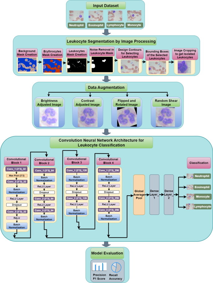
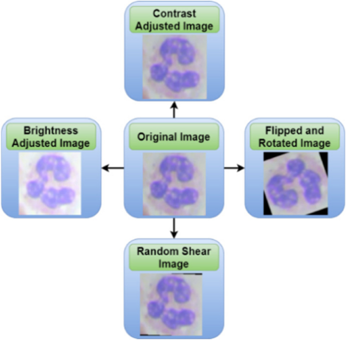
On this pipeline of the proposed work, leukocyte isolation by picture segmentation is carried out utilizing varied steps which can be mentioned within the following sections. Determine 1 shows the pipeline of the proposed work. The duty of robotically classifying leukocytes in blood photographs is difficult because of the variability of their look. The determine breaks down leukocyte classification into a number of phases. First, the enter dataset is preprocessed. The preprocessing workflow consists of three pivotal phases: the creation of Background Masks, Erythrocytes Masks, and Leukocytes Masks. Leukocyte segmentation is completed utilizing picture processing strategies, encompassing procedures reminiscent of background subtraction, noise elimination, and contouring. Leukocytes bear information augmentation methods, which contain changes in brightness and distinction, flipping, and random shearing. These augmentation processes purpose to reinforce the general adaptability and robustness of the CNN mannequin.
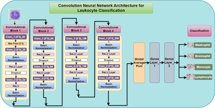
Subsequent, the preprocessed information is fed into the convolutional neural community. CNNs are a kind of synthetic neural community which can be well-suited for picture recognition duties. The CNN within the determine consists of 4 convolutional blocks, every containing convolutional layers, a max pooling layer, and a ReLU layer. Convolutional layers extract options from the info, max pooling layers cut back the dimensionality of the info, and ReLU layers introduce non-linearity. The stride worth for convolution and pooling layers is zero and no padding is utilized in these layers. After every convolutional block, there’s a batch normalization layer, which helps to enhance the coaching velocity and stability of the community.
Following the convolutional blocks, there are a number of totally related layers. These layers take the output from the convolutional blocks and map it to the ultimate class labels (neutrophil, eosinophil, lymphocyte, monocyte). Lastly, the mannequin is evaluated utilizing metrics reminiscent of precision, recall, F1 rating, and accuracy.
Total, the convolutional neural community structure within the determine gives a complete strategy to classifying leukocytes in blood photographs. By segmenting the leukocytes, preprocessing the info, and utilizing a CNN with acceptable layers, the mannequin can obtain excessive accuracy in classifying the several types of leukocytes. The proposed mannequin is analysed utilizing the Google Colab platform with Python.
Enter dataset
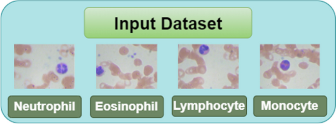
To validate the proposed mannequin, blood samples have been collected from a web-based supply. Blood pattern photographs are collected from a publicly out there dataset from Kaggle Mooney et al. [20]. The dataset comprises 4 leukocyte varieties named Neutrophil (NE), Eosinophil (EO), Lymphocyte (LM), and Monocyte (MN) as proven within the Fig. 2. It consists of a complete of 12,444 blood pattern photographs out of which 3144 photographs belong to EO class, 3139 to LM, 3132 to MN and 3171 to NE class. The splitting ratio of 80:20 is used for coaching and testing of the mannequin in line with which the whole rely of coaching photographs is 9955 and the rely of testing photographs is 2489. The dimensions of the enter picture is 256 * 192. Determine 2 exhibits the blood samples obtained from the Kaggle dataset.
Leukocyte isolation by picture segmentation
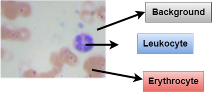
A blood cell picture consists of three components as proven in Fig. 3. First, the picture undergoes background subtraction to take away erythrocytes and different non-leukocyte components. Then, a noise elimination step cleans up the picture. Subsequent, the leukocytes are segmented from the background utilizing a masks creation course of. Lastly, the design corners are eliminated, abandoning the remoted leukocytes. Right here within the blood cell picture, grey half is the background whereas, the leukocyte is proven in blue coloration and the darkish brown half is the erythrocyte. For the segmentation of photographs and for isolating leukocytes, it’s essential to know the place of leukocytes within the coaching samples. For the detection of leukocytes, the processing is finished by making a masks for the background detection. After that, a masks is created for the detection of erythrocytes. Easy masks are created for filtering out the background space to extract leukocytes. Determine 4 exhibits the leukocyte isolation by picture segmentation.
Background masks creation
In blood cell photographs, the background space doesn’t include erythrocytes in addition to leukocytes and it’s grey in coloration. The grey pixel within the picture consists of the crimson, blue, and inexperienced elements within the majority. For creating background masks, threshold (threshold_1) is calculated utilizing Otsu thresholding methodology. So, the background masks is created utilizing the next Eq. (1)
$${textual content{Background}}_mathrm{masks }= ({textual content{img}}[:, :, 0] >mathrm{ threshold}_1) & ({textual content{img}}[:, :, 1] >mathrm{ threshold}_1) & ({textual content{img}}[:, :, 2] >mathrm{ threshold}_1)$$
(1)
On this equation, crimson element i.e. ({textual content{img}}[:, :, 0]), inexperienced element i.e. ({textual content{img}}[:, :, 1]) and blue element i.e. ({textual content{img}}[:, :, 2) all are having pixel value greater than threshold_1 whose value comes out to be 182 here. All background pixels satisfying the above equation are highlighted in blue color in Fig. 5.
Erythrocytes mask creation
In blood cell image shown in Fig. 3 erythrocyte is shown in light brown color. So, to extract the erythrocyte area from the blood cell images, the erythrocyte mask is created with a red component where the red pixel value is greater than the blue pixel value or the blue pixel value should be less than threshold value which comes out to be 150 calculated using Otsu thresholding method.
This function will detect the erythrocytes. So, the erythrocyte mask is created using the following Eq. (2)
$${text{Erythrocyte}}_mathrm{mask }= ({text{img}}[:, :, 2] <mathrm{ threshold}_2) | ({textual content{img}}[:, :, 0] >mathrm{ img}[:, :, 2])$$
(2)
On this equation, blue element i.e. ({textual content{img}}[:, :, 2]) will all the time be lower than crimson element i.e. ({textual content{img}}[:, :, 2]). Right here, blue element i.e. ({textual content{img}}[:, :, 2]) is lower than 150 or crimson element i.e ({textual content{img}}[:, :, 2]) is larger than blue element i.e. ({textual content{img}}[:, :, 2]). All erythrocyte pixels satisfying the above equation are highlighted in crimson coloration in Fig. 6.
Leukocytes masks creation
The leukocyte masks is created that doesn’t embrace a background masks and erythrocyte masks. So, the leukocyte masks is created utilizing the next Eq. (3)
$${textual content{Leukocyte}}_mathrm{masks }= sim {textual content{is}}_{textual content{background}}({textual content{img}}) & sim {textual content{is}}_{textual content{erytrocyte}}({textual content{img}})$$
(3)
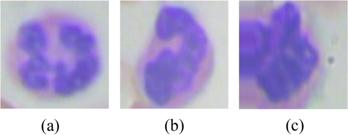
Determine 7 exhibits the picture samples with leukocyte masks. From Fig. 7 (a), (b), and (c) it may be seen that the leukocyte masks is proven in white coloration whereas, the background of the blood pattern picture is black.
Noise elimination in leukocyte masks
To take away the noise from the leukocyte masks morphological operations are used. From Fig. 7 proven within the final part, it may be seen that noise is current in leukocyte masks photographs. To do away with the little scraps and make the masks rounder, opening morphological operation is used i.e. erosion adopted by dilation. Determine 8 exhibits the eliminated noise photographs of leukocyte masks. The equations of dilation, erosion, and opening are given in Eqs. 4, 5, and 6 respectively. The equation of dilation operation is
$$P oplus Q=left{R|left[left(Q{wedge }_{z}right)cap Pright]epsilon Pright}$$
(4)
Right here, (P) is the picture and (Q) is the structuring ingredient. (left(Qwedge _zright)) it means to take a mirrored image of (Q) about its origin and shift it by (R). Due to this fact, dilation of (P) with (Q) is a set of all displacements, (R) such that (left(Qwedge _zright)) and (P) overlap by at the least one ingredient. The equation of abrasion operation is
$$P circleddash Q=leftleft(Qwedge _zright)epsilon Pright$$
(5)
Right here, the erosion of (P) by (Q) is a set of all factors that (Q), shifted by (R) is a subset of (P) that’s (Q) is completely contained inside (P). Erosion reduces the variety of pixels from the article boundary. The equation of opening operation is
$$P circ Q=OPENleft(P,Qright)=left(P circleddash Qright) oplus Q$$
(6)
Morphological opening of a picture is erosion adopted by dilation.
Bounding packing containers of the chosen leukocytes
On this step, the masks having the best space within the picture pattern is marked as leukocyte with the bounding packing containers and the define coloration is made crimson as proven in Fig. 9.
Picture cropping to get remoted leukocytes
On this step, the chosen leukocyte picture is cropped to a picture measurement of 128 * 128 as proven in Fig. 10.
Knowledge augmentation
Overfitting is a problem for giant neural networks in organic purposes since solely a comparatively small variety of datasets can be found. The dataset is subsequently expanded to attain better efficiency.
Numerous operations reminiscent of rotation, flipping, distinction, brightness, and random shear are utilized to every picture enter throughout information augmentation as proven in Fig. 11. Thereby rising the general coaching picture rely is an effective factor. Photographs are then divided into two units: one for coaching and one other for testing causes. The CNN mannequin is used to coach the mannequin.
Deep convolution neural community structure for segmented leukocyte
The convolutional neural community (CNN) structure within the Fig. 12 is a four-block deep studying mannequin designed to categorise leukocytes in blood smear photographs. Convolutional layer applies filters (discovered kernels) that slide throughout the enter picture, extracting options like edges and textures. The variety of filters will increase progressively via the blocks, permitting the community to check progressively complicated options. ReLU activation layer introduces non-linearity into the community, enabling it to study complicated relationships between options. Batch normalization layer reduces inside covariate shift, stabilizing the coaching course of and bettering the mannequin’s generalizability.
Following these convolutional blocks, a world common pooling layer captures spatial info from the characteristic maps, producing a fixed-size output that is appropriate for feeding into the ultimate densely related output layer. This last layer has 4 output neurons, akin to the 4 leukocyte lessons i.e. neutrophil, eosinophil, lymphocyte, and monocyte.
The primary convolutional block consists of three convolution layers with filter measurement of three * 3 and a complete of 32 filters, 2 batch normalization layers, 1 dropout, and 1 max pool layer with 2 * 2 filter measurement with 2 ReLU layers.
The second convolutional block comprises 3 convolution layers with filter measurement of three * 3 and a complete of 64 filters, 2 batch normalization layers, 1 dropout, and a couple of ReLU layers. The third convolutional block comprises two convolution layers with filter measurement of three * 3 and 128 whole filters, two batch normalization layers and two ReLU layers. The fourth convolutional block consists of three convolution layers, two batch normalization layers, two ReLU layers and one dropout layer. After the 4 convolutional blocks, the worldwide common pooling layer is connected with two dense layers. Within the final step, the blood pattern picture is assessed into the 4 lessons named as Neutrophil, Eosinophil, Monocyte and Lymphocyte. Total, this CNN structure employs a step-wise strategy, progressively extracting higher-level options from the enter picture to in the end obtain correct leukocyte classification.
The rationale behind utilizing a deep Convolutional Neural Community (CNN) for leukocyte classification lies in its capability to robotically study hierarchical options from uncooked information. Deep CNN has extra convolution layers to seize extra complicated options within the leukocyte photographs to study deep summary info of the info, doubtlessly main to higher efficiency. CNNs are well-suited for this job as a result of they’ll seize spatial hierarchies of options in photographs via their convolutional layers, which apply filters to detect patterns at totally different spatial scales. The proposed strategy has whole parameters as 134,853, with 134,085 as Trainable parameters and 768 as Non-trainable parameters.


