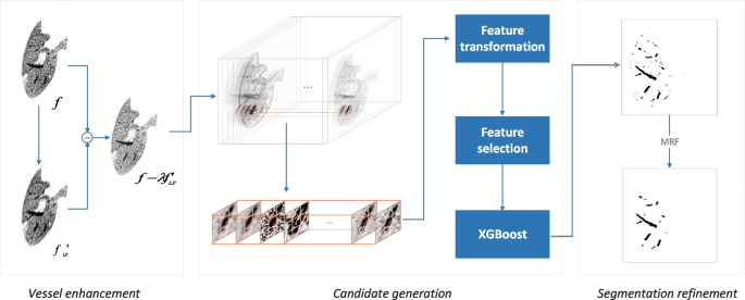
The proposed liver vessel segmentation mannequin composes of three modules: vessel enhancement, candidate technology, and segmentation refinement; see Fig. 2. The main points are as follows.
Diagram of the proposed liver vessel segmentation mannequin. It composes vessel enhancement, candidate technology, and segmentation refinement. Vessel enhancement is achieved by fading out the background however strengthening boundary areas, candidate vessels are obtained by XGBoost feeding with options generated from intensive picture filters, and refinement is fulfilled by a refined Markov random discipline
Vessel enhancement
Two procedures are utilized to the uncooked photos to reinforce the perimeters between vessel areas and different liver tissues, together with calibration and distinction.
Calibration is critical because the uncooked picture might have to be clipped to the suitable window for vessel evaluation. To this finish, we mechanically decide the window heart and width by a statistical method. Exactly, the imply (mu) and normal deviation (sigma) of vessel intensities are decided. Then, the intensities of all photos are clipped into an interval ([{mu } – 3{sigma }, {mu } + 3 {sigma }]). These clipped intensities are additional normalized to alleviate the systematic bias between completely different imaging gadgets by
$$start{aligned} f^{prime }(x,y)=alpha (f(x,y)-mu )/sigma )+c, finish{aligned}$$
the place f(x, y) is the preliminary depth of a picture at place (x, y), (alpha) and c are used to remodel the normalized values into grey scales from 0 to 255.
After calibration, the vessels are enhanced by
$$start{aligned} f^{prime }(x,y)=f(x,y)-lambda f(x,y)circledast okay(x,y), finish{aligned}$$
the place okay(x, y) is a kernel of a low-pass filter, (lambda) controls its magnitude, and (circledast) means convolution. This operation helps wash out many liver tissues and makes vessels stand out.
Candidate technology
Function transformation
The filters used to retrieve options from photos embrace CLAHE (distinction restricted adaptive histogram equalization) [23], Gabor filter [24], Gamma Correction [25], Gaussian filter [26], Hessian [7], Laplacian operator [27], Median filter [28], Imply filter [29], Minimal filter [30], Bilateral filter [31], Sobel operator [32], Canny edge detector [33], in addition to the ten filters predefined within the imageFilter module of Pillow [34], that are BLUR, CONTOUR, DETAIL, EDGE_ENHANCE, EDGE_ENHANCE_MORE, EMBOSS, FIND_EDGES, SMOOTH, SMOOTH_MORE and SHARPEN. The mathematical definitions of those filters/operators are proven in Desk 1.
These filters have their distinctive deserves in capturing options from photos. Thus, the data obtained on this means is enough to characterize vessels.
Context-aware vessel identification
Primarily based on the filters, every pixel is represented by a d-dimensional vector containing its authentic depth in addition to all of the values generated by the filters. Therefore, the context in addition to the vessel areas might be represented by a (ntimes d) vector with n the variety of neighbors surrounding the pixel to be labeled.
A pixel (F(i^{prime },j^{prime },okay^{prime })) is deemed as an h-hop neighbor of the curiosity pixel F(i, j, okay) if (textual content {min}(|i-i^{prime }|, |j-j^{prime }|, |k-k^{prime }|) le h), the place i, j and okay are the indices of a pixel, i and j are used to find the pixel in a slice, and okay is used to find the slice in a quantity. The h is about to 1, 2 and three, ensuing within the voxel dimension of (3times 3times 3), (5times 5times 5) and (7times 7times 7), respectively. For the 2D scenario, solely i and j are thought of.
The pixel in addition to its neighbors kind a voxel whose options are obtained from its constituent pixels, the place its label is the masks of the central pixel. The options are obtained through the use of the above filters. The ultimate options of the voxel are enter into XGBoost [21] for function choice and pixel classification.
Segmentation refinement
The vessel segmentation is additional refined by a Markov random discipline (MRF) [35] because the classification is simply carried out on pixel degree that ignores the correlation between pixels.
An MRF is a graph having ({G}=({V},{E})), the place V is the set of nodes (e.g., the pixels of a picture), and E is the perimeters connecting the nodes in V (e.g., the adjacency pixels). For a random variable (v_{i}) in G, the likelihood of (P(V=v_{i})) is unbiased of different variables given its neighbors (N(v_{i})) that’s named because the Markov blanket. That being mentioned,
$$start{aligned} P(V=v_{i}|V- v_{i})=P(V=v_{i}|N(v_{i}). finish{aligned}$$
Primarily based on the Hammersley-Clifford theorem [36], it may be expressed as
$$start{aligned} P(V=v_{i}|N(v_{i}))=frac{1}{Z}textual content {exp}(-E(V=v_{i}|N(v_{i}))), finish{aligned}$$
the place (E(cdot )) is an vitality operate and Z is the partition operate computed by (Z=sum _{v_{i}}E(v_{i})). On this examine (E(v_{i})) is calculated by
$$start{aligned} E(v_{i})= & {} E_{textual content {depth}}(v_{i})+lambda E_{textual content {gradient}}(v_{i}) = & {} sum rho (u_{i}-v_{i},sigma _{i}) + lambda sum limits _{v_{j}in N(v_{i})}rho (v_{i}-v_{j},sigma _{g}), finish{aligned}$$
the place (u_{i}) is the refined worth of the variable (v_{i}) and (rho (x,sigma )) is the Lorentzian operate [37] outlined by
$$start{aligned} rho (x,sigma )=log left( 1+left( frac{x}{sigma }proper) ^{2}/2 proper) . finish{aligned}$$
By minimizing the vitality operate E, we obtained the refined segmentation of the vessels based mostly on the pixel-wise classification outcomes.


